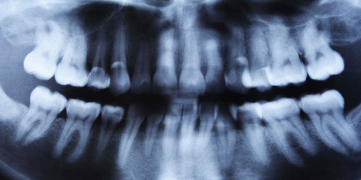
Good Dentistry Needs Good X-Rays

A type of radiation we call X-rays are used to take pictures (radiographs) of the jaw bone and teeth. They allow me to evaluate your mouth better for the presence of caries (decay) in the teeth and bone loss from periodontal disease. We also look for signs of pathology (abnormal changes)and A Periapical Xray showing the entire lower left molar teeth diseases such as oral cancers, sinus infections, gum diseases, and jaw joint (TMJ) problems. Many diseases that would go undetected for a long period of time if we rely only on visual examination. Many problems can be found at an early treatable stage through x-rays. Due to early detection, many diseases are treated without the need for drastic treatments, such as roots canals or extractions.
What are the types of Radiographs (xrays) that are taken?
Periapical radiographs give us a full view of the tooth crown, roots and surrounding bone. They are excellent for diagnosing periodontal disease and pathology (abnormalities) around the teeth. They silver fillings show up as bright white, most composite fillings are light gray, the enamel that covers the tooth is a slightly darker gray, dentin that supports the enamel and A Bite Wing Xray to detect caries (decay) between the teeth and the bone heightmakes up the root is a shade darker, bone is similar to dentin and the soft tissue of the pulp and gum tissue is the darker gray. Caries (decay) shows up as the darkest of grays, in most cases.
Bite Wing Radiographs are taken with you biting on an xray holder that positions the film half above and half below the biting plane. In that film, we can see just the crowns of the upper and lower teeth. It is primarily used to detect caries and get a sense of the periodontal bone level.
Both of the above radioLaptop Computer used to Record and Store the Xray Imagesgraphs can be taken with standard E-speed (fastest xray film speed available) or with an xray detector hooked up to a computer (which is called Computerized Digital Radiography, or CDR for short.) CDR has the advantage of using 1/5th (only 20%) of the xray dose as is needed to get the same image on E-speed film. With CDR, we also get the result in 7 Seconds rather than after 7 Minutes of processing.
Above is the Dell Latitude Notebook Computer with the CDR detector next to it. You can see the films on the computer screen and each can be enlarged to full screen.
Another type of radiograph we use in the office is the Panoramic Radiograph. We get a specialized picture of all of the teeth and both upper and lower jaws (Maxilla and Mandible) in one wide film. The Panoramic Radiograph is taken by a State of the Art Machine where the patient puts his chin on a rest and the machine moves the xray head (source) and the film around the patient’s head in about 7 Seconds.
Should I be concerned about the amount of Radiation?
Many patients ask about the exposure to x-ray radiation and possible health risks. I too have been concerned about this.
When I was doing research on the effects of radiation and chemotherapy on the growth and development of children’s teeth and jaws, I had work with the radiation physicists at Memorial Sloan Kettering Cancer and determine the dose for the diagnostic dental x -rays we used in the study.
On average, 21 conventional dental xrays taken with the E-speed film we use is equal to that which a person gets from while flying from New York to Denver or Miami. (Up at 30,000 feet the background cosmic radiation is higher due to thinner and less atmosphere above you.) In our office we use a specialized, selective 21-film periapical full mouth survey or a 4-film Bite Wing Survey to assess our patients teeth.
A combination of a panoramic x-ray machine and E-speed film to keep exposure to x-ray radiation at a minimum. Even the Panoramic x-ray machines take x-rays of a patient’s complete upper and lower jaw, sinuses, A typical Full Mouth Survey of Radiographsjaw joint, and teeth using approximately the same amount of radiation you’d get on a one-way plane trip to Denver. E-speed film allows x-rays to be taken 50% faster than conventional x-rays cutting exposure in half. Overall, it takes about 50 dental x-rays to equal the exposure of just one chest x-ray.
However, even with such low exposure it is our policy to take x-rays only when necessary and to keep your exposure to a minimum.
How often do you recommend taking xrays?
I have developed a specific set of guidelines for my patients.
Since New York State malpractice law requires (and you can not waive your rights for) me to do a complete oral diagnosis, you will need a recent full mouth series. If you have taken one within the last three years, please let us know and we will do everything we can to obtain it from your previous dentist with your permission.
Full mouth series are generally taken once in a patient’s 20’s and 30’s and every 3 to 5 years thereafter, due to the increased incidence of periodontal bone changes as an indicator of periodontal disease.
Bite wing xrays (4 for adults and 2 for children) are taken on a schedule that matches your decay experience.
Patients with active decay will have one taken in 6 months to see if new areas of decay are present.
If you have no active decay, but have previous decay between the teeth, we take the bite wing xrays once a year (but still see you every 6 months for examination and cleaning.)
Patients who have no or very few fillings between teeth will only get bite wings every 18 months.
Most (about 75%) of my patients are on 12 or 18 schedules of Bite Wing Xray Surveys.
What if I need the x-rays to see another dentist?
The original radiographs can always be send to any other dentist, due to systems I set up because I have many patients who are here in New York for limited time. This includes a large number of the residents and fellows doing training at New York Hospital, Memorial Sloan-Kettering Cancer Center, HSS, Rockefeller Univ, etc. Others patients work for multinational companies and relocate out of NY after receiving care in my office. For these reasons, all intra-oral films (those taken in the mouth) are taken with 2-film packs (there are 2 x-ray films in each of the small plastic-covered holders) that give us two identical films for each x-ray exposure. The CDR (Computerized Digital Radiography) system can printout as many copies as we need and the images can be sent electronically via phone or internet to any other dentist with a computer. Panoramic films can be duplicated by us in the office.
If you have any questions or concerns about x-ray examinations, please feel free to call the office.
