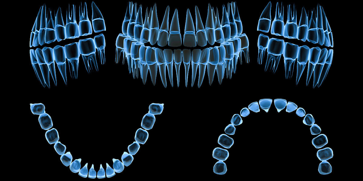
Computerized Digital Radiography

I brought Schick CDR into the office in October, 1998. It has revolutionized how I diagnose emergency pain patients and how I do endodontics (root canal therapy). Patients who want x-rays taken with 20% of the usual radiation dose for conventional dental film x-rays can also have CDR full mouth x-ray series and 4 Bite Wing (Cavity Checking) x-ray series.
Our Schick Computerized Digital Radiography (CDR) System:
- Is the current State of the Art technology.
- Reduces the radiation to 80% less than conventional x-ray films.
- Reduces the time (7 seconds) from exposure to viewing the image.
Further benefits of the CDR System are as follows:
- I have the extra time to discuss what we see and what treatment options are recommended, instead of waiting 7 minutes for dark room processing.
- All images are accessible and linked to the Dental Software.
- Images can be magnified to full screen.
- Accurate measurements (to tenths of mm’s) are easily done, which is helpful with endodontics, implants and bone loss evaluation.
- The contrast and darkness can be modified for better viewing.
- The images can be colorized for easier viewing and interpretation.
- We have instant retrieval of all CDR x-ray images.
- We can put up to 4 images on the screen for comparison of radiographs.
- You can see what I am seeing with the images magnified many times larger.
- We are reducing the about of chemicals that need to be discarded or processed to prevent pollution.
- Instant retakes can be made if more information is needed with minimal radiation
- We can get rapid consultation by our specialists. Recently, while treating a tough endodontic case, I had a question in my mind of the best way to treat a situation. I sent the CDR image to be reviewed by our endodontist across town. He looked at it on his computer screen at the same time as I did and discussed how he recommended that I proceed. He was also able to confirm that no referral of the patient was needed.
- The digital radiographs can be displayed, stored, printed, and sent anywhere via modem.
- Electronic claims processing should be available soon, so that we can attach the image file to the insurance claim file and send it electronically to the insurance company.
- Future advances potential (three dimensional radiography, second generation digital subtraction, teleradiography, etc.)
- The sensor used is 5 mm thick and the same size as regular dental films. The sensor is hard wired directly to the computer that generates and displays the image.
- We can ZOOM in on the area we need a closer look at.
- A Full Mouth Series of 18 to 21 Images can be taken and then each image enlarged and adjusted to see more features of the tooth, bone and surrounding structures.
